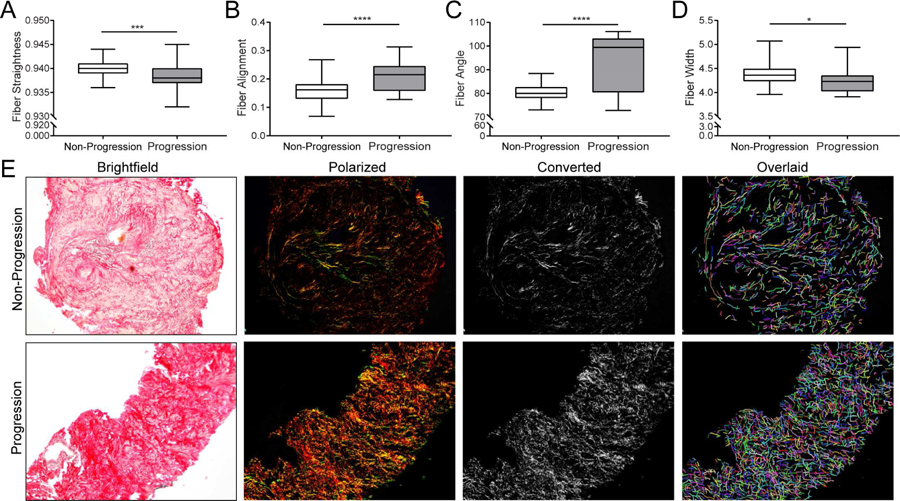
Figure 1.

Collagen Fibril Structure of Transition Zone Biopsy Specimens. Box and whisker plots indicating lower extreme, lower quartile, median (line), mean (+), upper quartile and upper extremes. A. Boxplot of data for collagen fiber linearity, or straightness, for which TZ biopsies were significantly less linear (i.e. wavy) for participants who experienced clinical progression than non-progression (p<0.001). Assessment of collagen fiber linearity or straightness was accomplished by measuring the distance of a straight line drawn between fiber end points divided by the distance of a line drawn along the entire fiber. Scores = 1.0 indicated perfect linearity, or fiber straightness, whereas scores <1.0 indicated fiber curvature. B. Boxplot of data for collagen fiber alignment indicates TZ biopsies from patients who experienced clinical progression had significantly increased fiber alignment than those who did not (p<0.0001). Collagen fiber alignment quantifies how the fibers are aligned with each other by measuring the similarity of all fiber angles. Scores = 1.0 indicated perfect fiber alignment with each other, whereas scores <1.0 indicated fibers that are less aligned with each other. C. Boxplot of data for collagen fiber angle indicates those that biopsies from men who experienced clinical progression had significantly larger angles than from those who did not (p<0.0001). Assessment of collagen fiber angle was accomplished by measuring the angle made between a straight line drawn between fiber end points and a horizontal line drawn across the fiber. D. Boxplot of data for collagen fiber width showing patients that biopsies from men who experienced clinical progression had thinner fibers than from those who did not (p<0.05). E. Representative picrosirius red stained brightfield, circular polarized, converted grayscale and CT-FIRE overlaid pseudo-colored fibers of core biopsies demonstrating collagen fiber alignment was more dense, thin, wavy and organized in TZ biopsy tissue from men treated with doxazosin + finasteride who experienced clinical progression (lower panel) compared to those who did not (upper panel).
