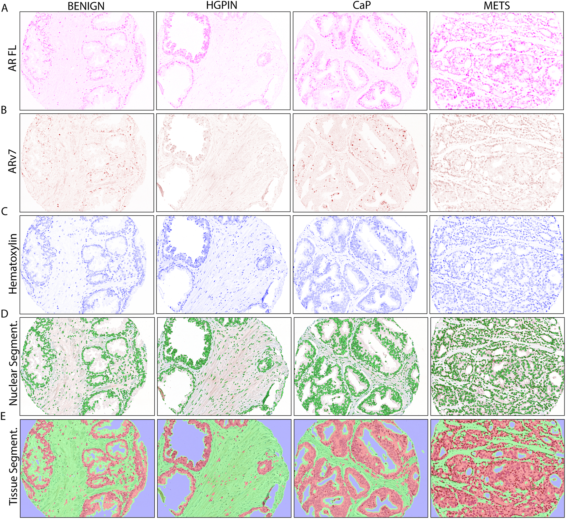
FIGURE 1. AR and ARv7 in Multiplexed IHC with Cell and Tissue Segmentation.

A. Optically isolated full-length AR (AR FL) staining (purple) in PRCA progression.
B. Optically isolated ARv7 staining (brown) in PRCA progression.
C. Optically isolated hematoxylin staining (blue) in PRCA progression.
D. Automated nuclear segmentation (green) in PRCA progression.
E. Automated tissue segmentation (epithelium=red, stroma=green) in PRCA progression.
BENIGN = benign prostatic tissue, n=52; HGPIN = high grade prostatic neoplasia, n=25; PRCA = prostate cancer, n=73; METS = metastasis, n=22. IHC image magnification = 20X.
