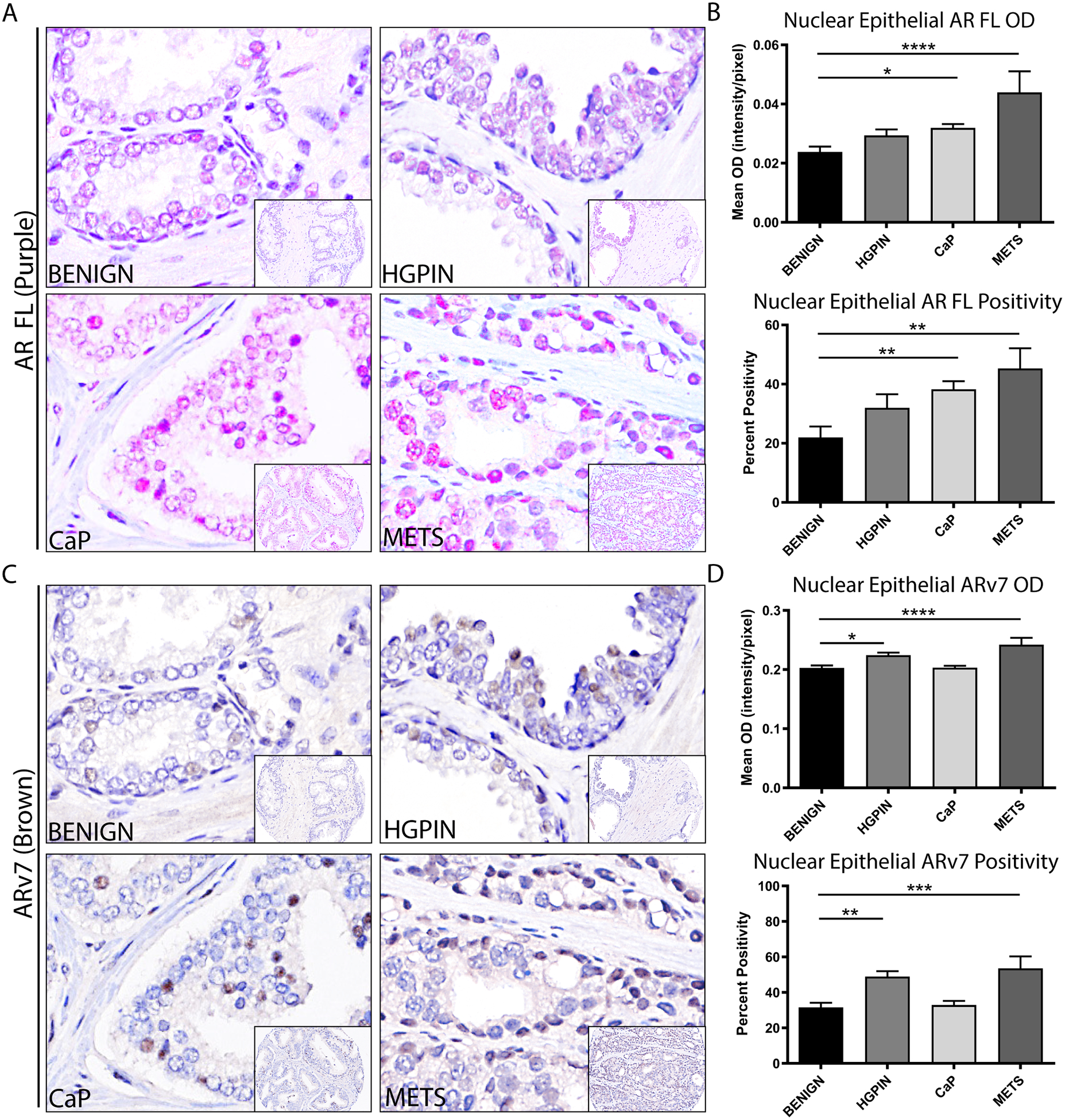
FIGURE 2. Independent AR and ARv7 Expression in PRCA Progression.

A. Representative images of full-length AR (AR FL, purple) with hematoxylin (blue), showing nuclear localization at all stages of PRCA progression.
B. Quantification of nuclear epithelial full-length AR (AR FL) in PRCA progression by mean optical density (top) and percent positivity (bottom) showed a significant increase in AR in PRCA and METS compared to BENIGN (p<0.05, p<0.0001 respectively for mean OD; p<0.01 for percent positivity).
C. Representative images of ARv7 (brown) with hematoxylin (blue), showing nuclear localization at all stages of PRCA progression.
D. Quantification of nuclear epithelial ARv7 in PRCA progression by mean optical density (top) and percent positivity (bottom) showed a significant increase in ARv7 in HGPIN and METS compared to BENIGN (p<0.05, p<0.0001 respectively for mean OD; p<0.01, p<0.001 respectively for percent positivity).
Image magnification = 100X with 20X inset.
