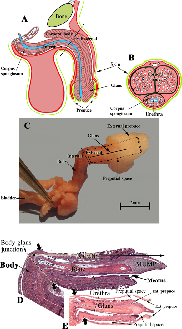
Figure 2.
Adult human and mouse penile anatomy. (A) Drawing of human penis in mid-sagittal view and (B) in transverse section. Note junction of the pendulous external portion and the internal portion of the human penis indicated by the dotted line in (A). (C) Photograph of a dissected adult mouse penis. The junction between the internal and external portions of the mouse penis occurs at the right-angle bend denoted by the dotted line and associated labels, “internal” and “external”. The forceps hold one of the bilateral crura. The internal portion contains the body of the mouse penis, while the external portion is called the glans. The glans in (C) cannot be seen because it lies within the preputial space (dotted lines); the position of the glans is indicated by . (D) Mid- sagittal hematoxylin–eosin stained section of the adult mouse penis with the external prepuce removed. (E) The mouse glans penis within the proximal portion of the external prepuce. Note reflection of epithelium of the external prepuce onto the surface of the penis indicated by the large solid arrows in both (D & E). (Adapted from Rodriguez et al. 2011 and Cunha et al. 2015 with permission).

