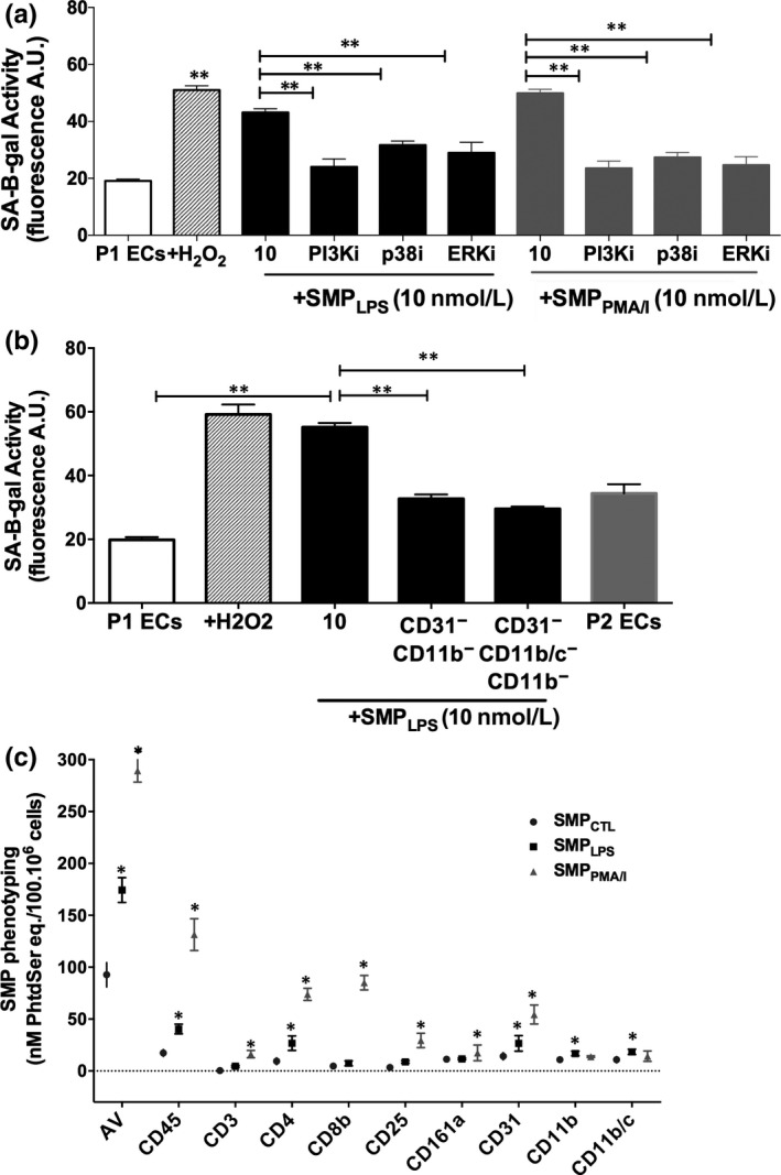
FIGURE 7.

SMPLPS induce endothelial senescence through MAP‐Kinase and PI3 kinase pathways, and via neutrophil‐derived MPs. A, P1ECs were incubated either with 100 μmol/L H2O2, or a selective inhibitor of PI3 kinase (PI3Ki, LY294002, 10 μmol/L), p38 MAPK (p38i, SB203580, 10 μmol/L) or ERK ½ (ERKi, PD98059, 10 μmol/L) for 1 h prior to the addition of 10 nmol/L SMPLPS or SMPPMA/I for 48 h before the determination of SA‐β‐gal activity. B, Effect of the removal of either CD31+/CD11b+‐MPs or CD31+/CD11b+/CD11b/c+‐ MPs on the endothelial pro‐senescent effect of SMPLPS. P1ECs were incubated with 10 nmol/L of SMPLPS and with non‐depleted suspensions for 48 h before the determination of SA‐β‐gal activity. C, Characterization of the cell origin of SMP shed from splenocytes stimulated by LPS or PMA/calcium ionophore. After 24‐h stimulation, SMPs were measured in cell supernatants by pro‐thrombinase assay and expressed as nmol/L PhtdSer per 100.106 cells. Characterization of the SMP cell origin was performed by capturing SMPs onto biotinylated antibodies directed against leucocyte CDs before quantification by pro‐thrombinase assay. Data are expressed as mean ± SEM of experiments performed at least on three different cell cultures. AV: Annexin V. *P < .05, **P < .01
