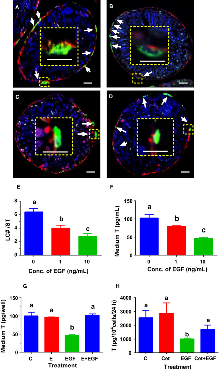
FIGURE 2.

Effects of EGF on differentiation of stem Leydig cells (SLCs) in vitro. Leydig cell (LC)‐depleted seminiferous tubules (STs) were cultured LC differentiation medium (LDM) containing EGF (0 ng/mL, Panel A), EGF antagonist (erlotinib, E, 1 µmol/L, Panel B), EGF (10 ng/mL, Panel C) and EGF (10 ng/mL) + E (1 µmol/L, Panel D) for 14 d. LCs were identified by biomarker 11β‐hydroxysteroid dehydrogenase 1 (HSD11B1, green colour in the cytoplasm, white arrow). Peritubular myoid cells were stained by smooth muscle actin (red colour in the cytoplasm), drawing a boundary for the ST. DAPI serves as counterstaining. Inset is the magnified picture, showing that HSD11B1 positive LCs existed outside of STs. Bar = 10 μm. The HSD11B1‐positive LCs per ST in the cross section were presented in Panel E (mean ± SEM, n = 4). In Panel F, STs were cultured in LDM without or with 1 or 10 ng/mL EGF for 14 d, and medium T levels were measured (mean ± SEM, n = 4). In Panel G, STs were treated in LDM with 0 ng/kg EGF (control, C) or 100 nmol/L EGF antagonist (erlotinib, E), or 10 ng/mL EGF (EGF) or 100 nmol/L E plus 10 ng/mL EGF (E + EGF) for 14 d, and medium T levels were measured (mean ± SEM, n = 4). In Panel H, CD90+ SLCs were treated with 0 ng/kg EGF (control, C) or 5 μg/mL EGF antagonist (Cet), or 10 ng/mL EGF (EGF) or 5 μg/mL Cet plus 10 ng/mL EGF (Cet + EGF) for 14 d to produce T (mean ± SEM, n = 4). Identical letters designate no significant difference between two groups at P < .05
