
Figure 3.
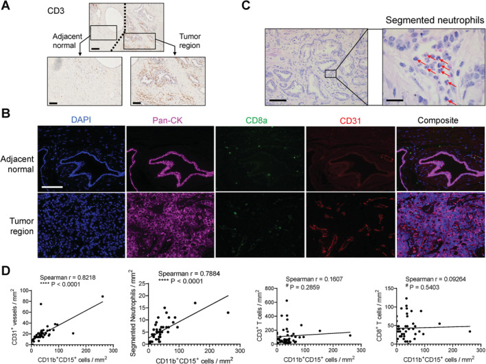
Positive correlation of tumor stromal PMN‐MDSCs with vascularization and segmented neutrophils, evaluated using primary tumor samples (N = 46, SBMF). (A) Representative IHC images of CD3. Scale bar, 200 μm (top) and 100 μm (bottom). (B) Representative images of primary tumor stained with the CD31/CD8a/pan‐CK/DAPI mIF panel. Scale bar, 100 μm. (C) Representative images of visually recognized segmented neutrophils (red arrows). Scale bar, 100 μm (left) and 20 μm (right). (D) Correlation analysis of the numbers of tumor stromal PMN‐MDSCs with CD31+ vessels, segmented neutrophils, CD3+ total T cells, and CD8+ cytotoxic T cells, tested by two‐tailed Spearman test.