
Figure 2.
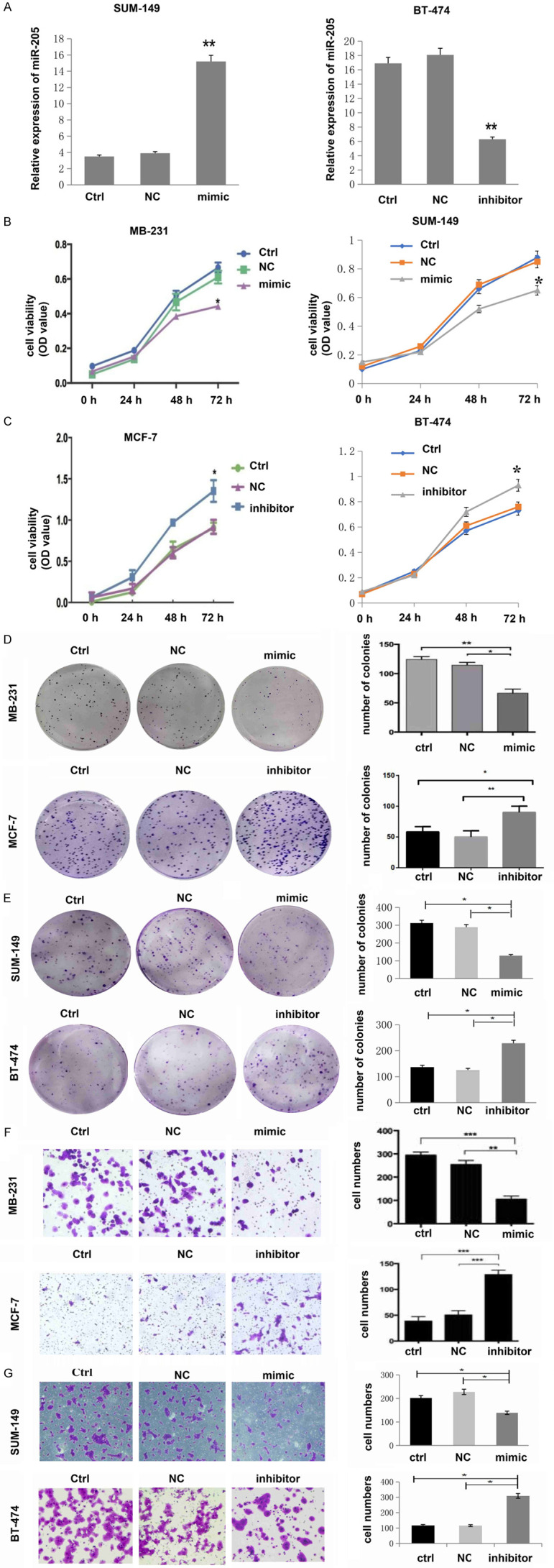
miR-205 is negatively associated with the malignant level of breast cancer cells. A. RT-PCR was used to detect the relative expression of miR-205 in cells transfected with miR-205 mimic or miR-205 inhibitor. **P<0.01, compared to Ctrl and NC. Data analysis was performed by an unpaired Student’s t-test. B. Cell proliferation was decreased after upregulation of miR-205 by infection with miR-205 mimic in MDA-MB-231 and SUM-149 cells (*P<0.05). C. Cell proliferation was increased after downregulation of miR-205 by infection with miR-205 inhibitor in MCF-7 and BT-474 cells (*P<0.05). D, E. Cell colony numbers were decreased after upregulation of miR-205 by infection with miR-205 mimic in MDA-MB-231 and SUM-149 cells, while they increased after downregulation of miR-205 by infection with a miR-205 inhibitor in MCF-7 cells and BT-474 cells (*P<0.05, **P<0.01). F, G. Cell migration ability was decreased after upregulation of miR-205 by infection with miR-205 mimics. However, the expression of miR-205 was increased after downregulation of miR-205 by infection with the miR-205 inhibitor (*P<0.05, **P<0.01, ***P<0.001). Each experiment was repeated three times, and the results are presented as the mean ± SD.