
Figure 6.
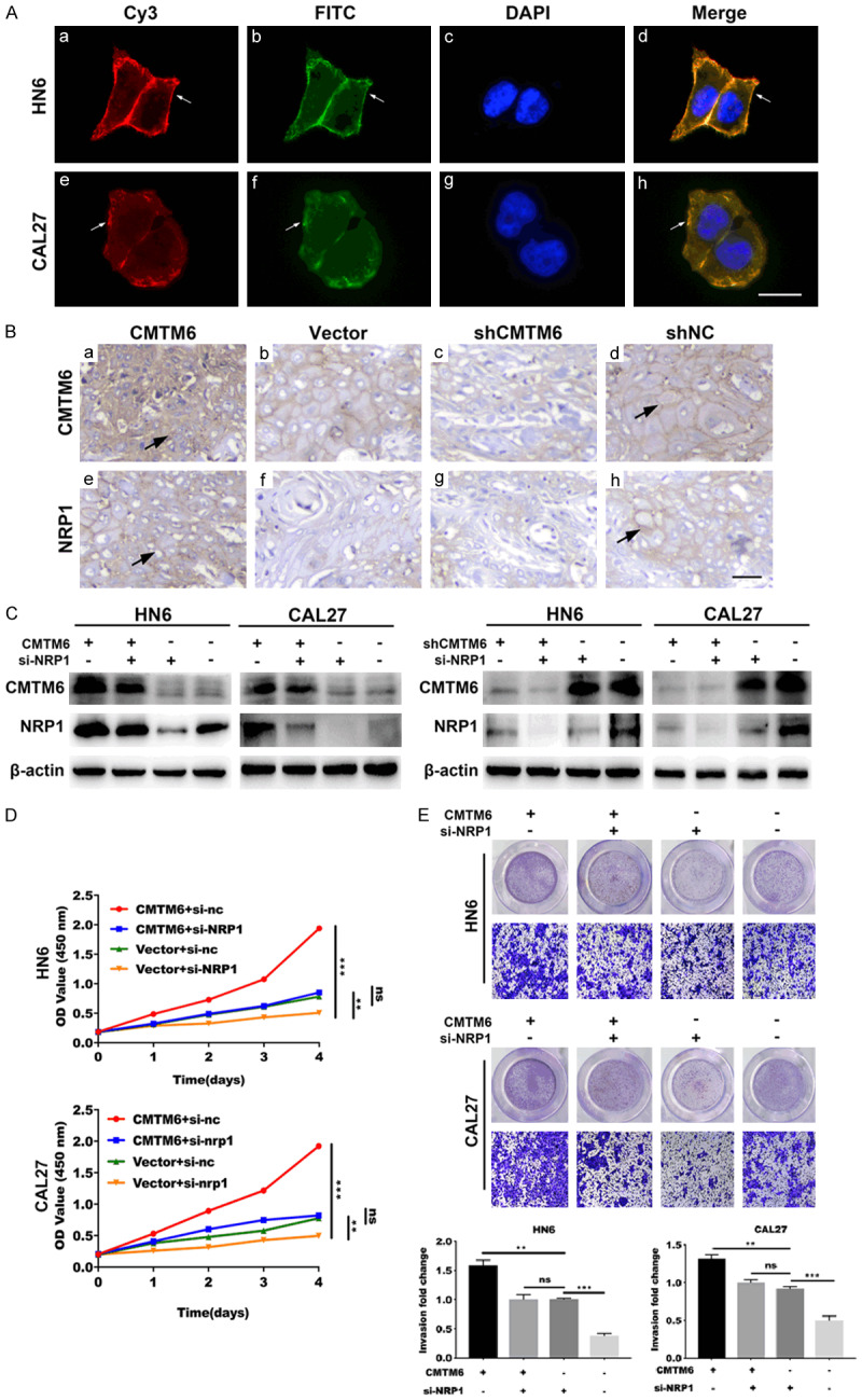
NRP1 is essential for CMTM6-mediated OSCC proliferation and invasion. (A) Confocal microscopy was utilized to capture the subcellular localization of CMTM6 (red) and NRP1 (green) expression in HN6 and CAL27 cells, followed by DAPI nuclear counterstaining (blue). Scale bar, 20 μm. (B) Mice xenograft tumors were analyzed by IHC to detect the co-expression of CMTM6 (a-d) and NRP1 (e-h). Overexpression of CMTM6 (a) resulted in higher NRP1 (e) protein staining. Scale bar, 50 μm. (C) Western blotting analysis was utilized to assess the expression of NRP1 in CMTM6 overexpressed or knockdown cells with the transfection of si-NRP1. (D) CCK-8 assay showed that the depletion of NRP1 significantly reduced the cell proliferation in CMTM6-overexpressed cells. (E) Transwell assay showed an increased invasive ability in CMTM6-transfected cells and the ability was restrained by NRP1 knockdown. Magnification: × 100. *P < 0.05, **P < 0.01, ***P < 0.001.