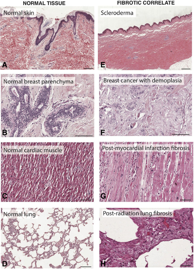
Fig. 3.

Organ fibrosis throughout the body. Representative histology of healthy tissue from the skin (A), breast (B), heart (C), and lung (D), compared with fibrotic tissue from those organs. Fibrotic tissue histology (E–H) demonstrates typical “hallmarks” of fibrosis, including densely aligned ECM fibers, decreased cellularity, and altered tissue architecture. Scale bars, 200 μm. Individual histology images were obtained from the Pathology Education Instructional Resource (PEIR) Digital Library and used with permission from Dr. Peter Anderson.
