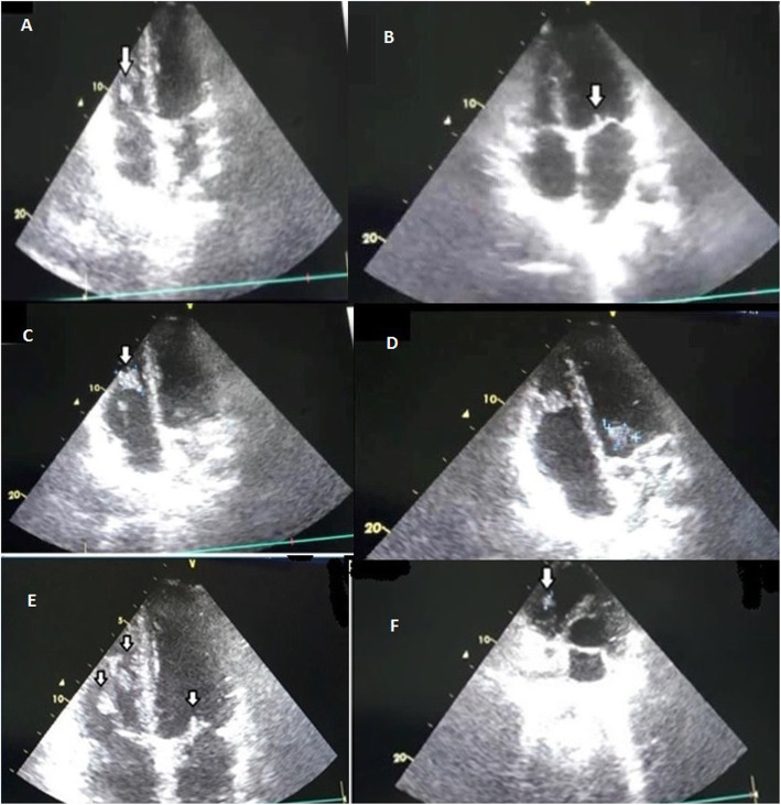
Fig. 3.
Transthoracic echocardiography (TTE): a four-chamber view showing a large-sized vegetation (arrow) seen on the right ventricular side of the leaflet of the tricuspid valve (TV); b four-chamber view showing a vegetation (arrow) on the ventricular side of the anterior leaflet of the mitral valve; c four-chamber view showing a large vegetation (arrow) on the lateral wall of the right ventricle; d Parasternal long axis view showing two large vegetations on mitral valve and lateral wall of RV; e Parasternal long axis view showing three large vegetations on mitral and tricuspid valves and lateral wall of RV; f Parasternal short-axis view showing a large vegetation on the ventricular side of the leaflet of TV

