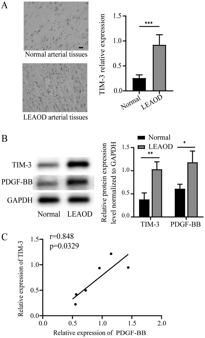
Figure 1.
Differences in TIM-3 expression between LEAOD and normal arterial tissues. (A) Immunohistochemical analysis of TIM-3 expression in LEAOD and normal arterial tissues (magnification, ×400). (B) TIM-2 and PDGF-BB protein expression levels in LEAOD and normal arterial tissues were determined by western blotting and quantification. (C) Correlation between the relative expression of PDGF-BB and TIM-3. *P<0.05, **P<0.01, ***P<0.001. TIM-3, T-cell immunoglobulin and mucin domain 3; LEAOD, lower extremity arteriosclerosis obliterans disease; PDGF-BB, platelet-derived growth factor-BB.

