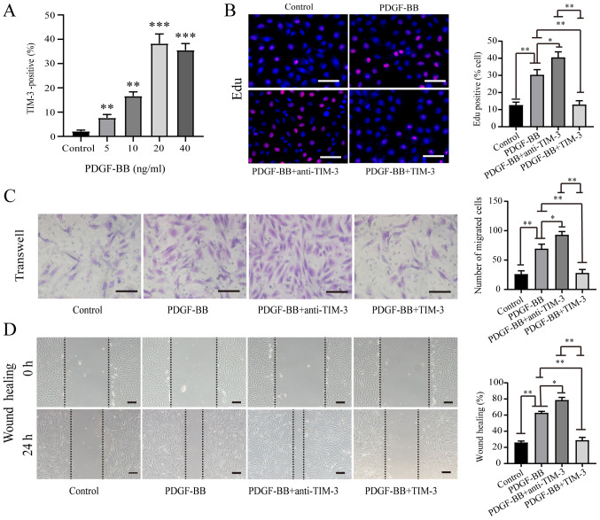
Figure 2.
TIM-3 inhibits PDGF-BB-induced proliferation and migration in HASMCs. (A) Quantification of flow cytometric analysis of the proportion of TIM-3+ PDGF-BB-stimulated HASMCs. **P<0.01 and ***P<0.001 vs. control group (B) EdU, (C) Transwell and (D) wound healing assays were used to assess proliferation and migration in PDGF-BB-stimulated (20 ng/ml) HASMCs in the presence or absence of TIM-3 (1,000 ng/ml) and anti-TIM-3 (10 µg/ml; n=5; scale bar, 100 µm). *P<0.05, **P<0.01. TIM-3, T-cell immunoglobulin and mucin domain 3; PDGF-BB, platelet-derived growth factor-BB; HASMCs, human artery vascular smooth muscle cells.

