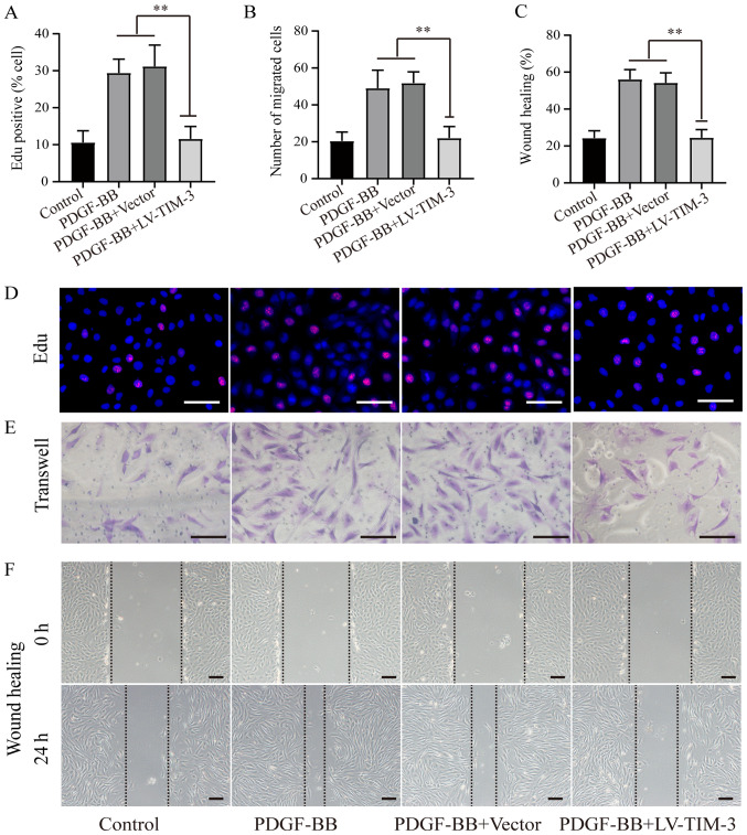
Figure 3.
Effect of TIM-3 overexpression on proliferation and migration in PDGF-BB-stimulated HASMCs. (A) Proportion of EdU-positive cells. (B) Number of migratory cells. (C) Ratio of wound healing. (D) EdU, (E) Transwell and (F) wound healing assays were performed to assess proliferation and migration in PDGF-BB-stimulated (20 ng/ml) HASMCs in the presence or absence of LV-TIM-3 (n=5; scale bar, 100 µm). **P<0.01 as indicated. TIM-3, T-cell immunoglobulin and mucin domain 3; PDGF-BB, platelet-derived growth factor-BB; HASMCs, human artery vascular smooth muscle cells; LV, lentivirus.

