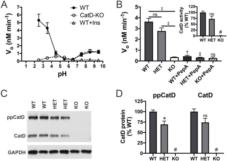
Fig. 1.
CatD activity and protein levels in brain extracts. a Aβ degradation in soluble brain extracts from 15-day-old WT and CatD-KO mice as a function of pH. Note that the abundant Aβ-degrading activity occurring at acidic pH is essentially absent in CatD-KO extracts. Note also that the smaller peak at neutral pH is inhibited by insulin (Ins), reflecting IDE activity [28]. Data are mean ± SEM for 5 replicates. †p < 0.01. b Aβ-degrading activity in extracts from 15-day-old CatD-KO, CatD-HET, and CatD-WT mice at pH 4.0. Note that the activity in WT and CatD-HET extracts is largely inhibited by the CatD inhibitor, pepstatin A (PepA). Data are mean ± SEM for 4 replicates. †p < 0.01; ‡p < 0.001; #p < 0.0001. Inset: CatD activity in brain extracts from WT, CatD-HET, and CatD-KO mice measured directly using a selective substrate. Data are mean ± SEM for 4 replicates.; #p < 0.0001. Note also that Aβ-degrading activity in the CatD-HET extracts is not reduced by 50% as expected from deletion of one of two CTSD alleles. c, d Representative western blot (c) and quantification of multiple samples (d) showing relative CatD levels in CatD-KO, CatD-HET, and CatD-WT mice. Note that, consistent with the activity data in b, CatD levels in CatD-HET brains are not 50% of those in WT brains. Data in d are mean ± SEM for 6 samples per genotype. *p < 0.05; #p < 0.0001

