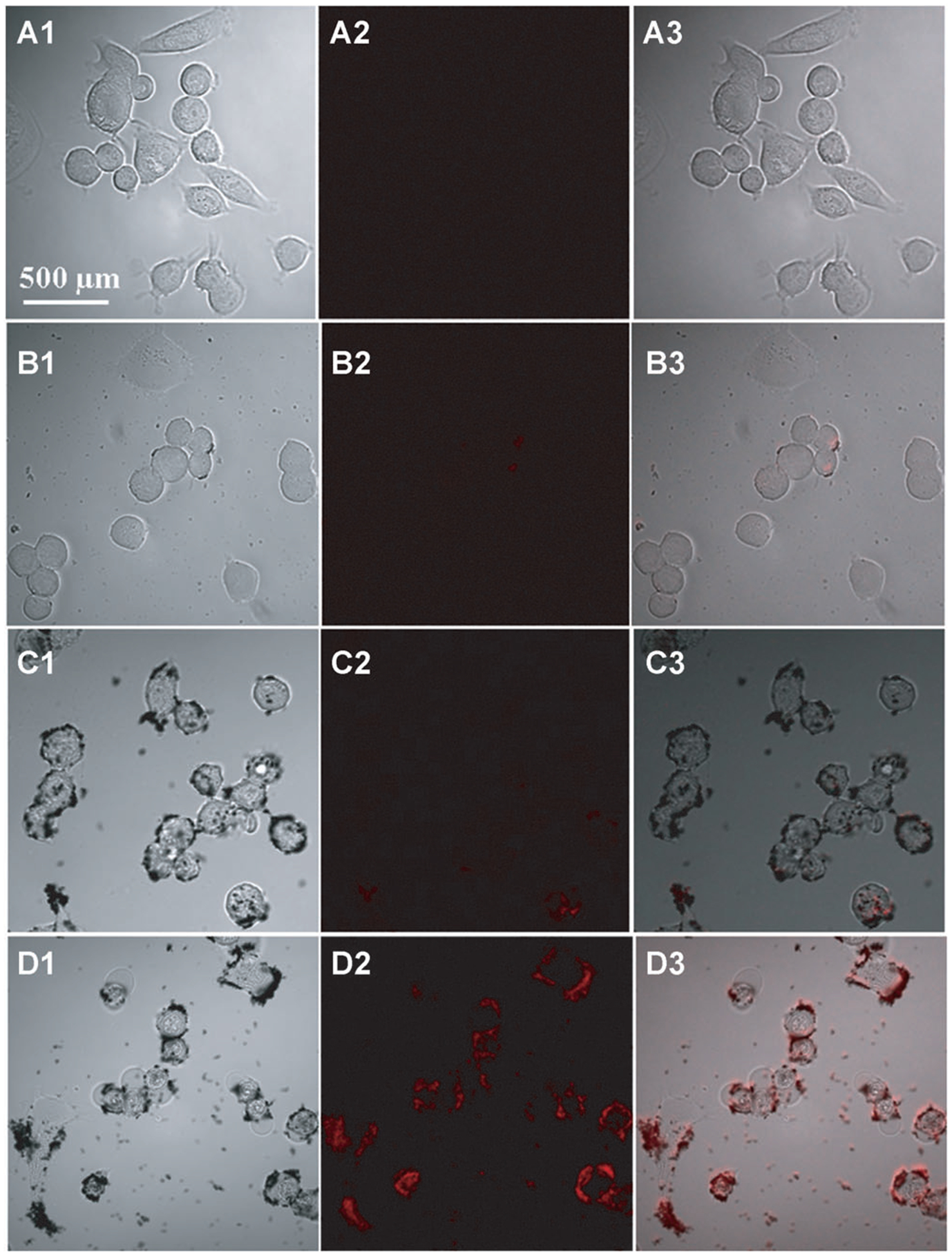
Figure 6.

Confocal fluorescence microscopy images of passively targeted SKBR3 cells with LS288-adsorbed AuNR 1, AuNR 2, and AuNR 4 collected using 785 nm excitation. The columns 1, 2, and 3 are bright field, fluorescence, and merged images of SKBR3 cells, respectively. The microscopy images in rows A, B, C, and D correspond to the dye alone, AuNR 1, AuNR 2, and AuNR 4.5, respectively.
