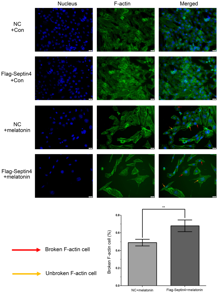
Figure 4.
Overexpression of Septin4 increases cytoskeleton destruction in hFOB 1.19 cells treated with a high melatonin concentration. hFOB 1.19 cells were untreated or treated with 4 mmol/l melatonin for 48 h. F-actin was visualized using phalloidin and the nucleus was stained using DAPI. The images were captured using fluorescence microscopy (×20) and an image of the two stains merged was shown. The yellow arrow represents the F-actin cytoskeleton and the red arrow depicts the destroyed cytoskeleton. The histogram indicates the percentage of cytoskeleton-destroyed cells among the total cells. **P<0.01, vs. NC group. GRP, glucose-regulated protein; NC, negative control; F-actin, fibrillar actin.

