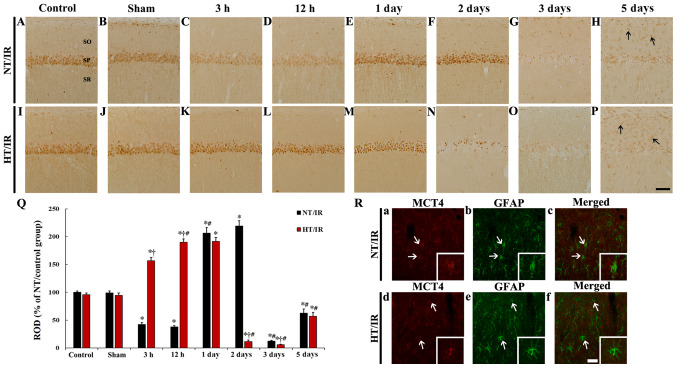
Figure 4.
MCT4 immunohistochemistry in CA1 of the (A) NT/control, (B) NT/sham, NT/IR at (C) 3 h, (D) 12 h, (E) 1 day, (F) 2 days, (G) 3 days and (H) 5 days after IR, (I) HT/control, (J) HT/sham and HT/IR groups at (K) 3 h, (L) 12 h, (M) 1 day, (N) 2 days, (O) 3 days and (P) 5 days after IR. In the NT/IR group, MCT4 immunoreactivity in the SP was markedly reduced at 3 and 12 h post-IR, significantly increased at 1 and 2 days post-IR, and barely observed at 3 and 5 days post-IR. In the HT/IR group, MCT4 immunoreactivity in the SP was significantly increased from 3 h post-IR, increased until 1 day post-IR, dramatically decreased at 2 days post-IR, and barely observed at 3 and 5 days post-IR. Note that MCT4 immunoreactivity was observed in non-pyramidal cells (arrows) in SO and SR at 5 days post-IR in both NT/IR and HT/IR groups. Scale bar, 50 µm. (Q) ROD of MCT4 immunoreactivity as % in CA1. *P<0.0001, significantly different from the NT/sham or HT/sham group; †P<0.0001, significantly different from the corresponding NT group; #P<0.0001 vs. pre-time point group. (R) Double immunofluorescence staining for MCT4 (red, a and d), GFAP (green, b and e), and merged (c and f) images at 5 days post-IR in the NT/IR (upper panels) and the HT/IR (lower panels) groups. MCT4 immunoreactivity is merged with GFAP immunoreactive astrocytes (arrows). Scale bar, 50 µm. CA1, cornu ammonis 1; IR, ischemia/reperfusion; NT, normothermia; HT, hyperthermia; MCT4, Monocarboxylate transporter 4; SP, stratum pyramidale; SO, strata oriens; SR, stratum radiatum; ROD, Relative optical density.

