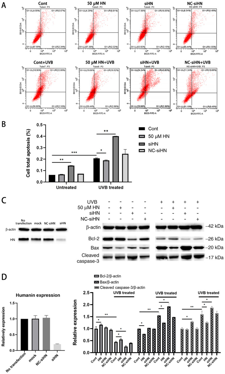
Figure 5.
Apoptosis assay of HLECs under oxidative stress induced by UVB. After pretreatment with 50 µM HN for 2 h or transfection with siRNA for 48 h, HLECs were subjected to 30 mJ/cm2 UVB radiation. (A) Annexin V/PI staining detected by flow cytometry. (B) Quantification of flow cytometry results. (C) Western blotting and (D) semi-quantification of the expression levels of the apoptosis-related proteins Bcl-2, Bax and cleaved caspase-3. Data are presented as the mean ± SD, n=3. *P<0.05, **P<0.01, ***P<0.001. HN, humanin; HLECs, human lens epithelial cells; UVB, type B UV; siHN, HN siRNA; siRNA, small interfering RNA; NC, negative control; Cont, control.

