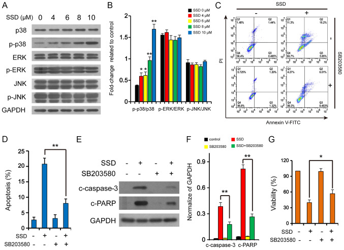
Figure 2.
SSD activates the p38 mitogen-activated protein kinase signaling pathway. (A) MDA-MB-231 cells were exposed to SSD for 24 h and the expression levels of JNK, p-JNK, ERK, p-ERK, p38 and p-p38 were detected by western blotting (n=3). (B) Semi-quantitative analysis of the expression levels from part A. *P<0.05, **P<0.01 vs. 0 µM. MDA-MB-231 cells were pretreated with 10 µM SB203580 for 2 h and then exposed to 10 µM SSD for 24 h. (C) Apoptotic cells were determined using flow cytometry (n=3). (D) Quantitative analysis of the apoptotic rate. **P<0.01. (E) Expression levels of c-caspase-3 and c-PARP were detected using western blotting. (F) Semi-quantitative analysis of the expression levels in part E. **P<0.01 vs. control. (G) Cell viability was determined using MTT assays (n=3). *P<0.05. Data are presented as the mean ± SD. SSD, saikosaponin D; c-, cleaved; p-, phosphorylated; PI, propidium iodide; PARP, poly (ADP-ribose) polymerase.

