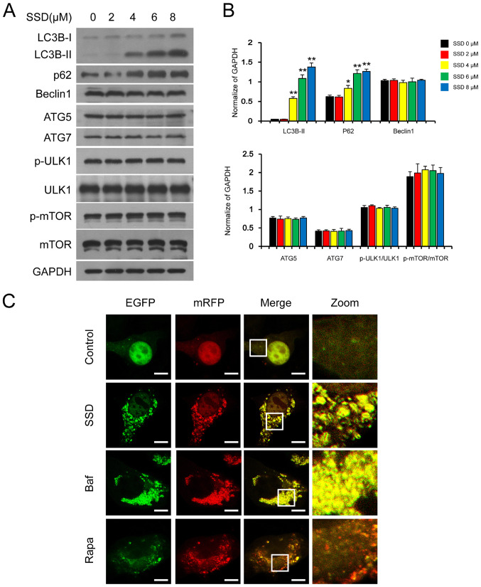
Figure 3.
SSD inhibits autophagic degradation. (A) MDA-MB-231 cells were exposed to SSD for 24 h and the expression levels of LC3, p62, Beclin 1, p-mTOR, mTOR, ATG7, ATG5, p-ULK1 and ULK1 were analyzed using western blotting. (B) Semi-quantitative analysis of the expression levels of part A. n=3; *P<0.05, **P<0.01 vs. 0 µM. (C) Cells were transfected with tfLC3 and exposed to 8 µM SSD, 0.25 µM Rapa or 20 nM Baf for 24 h. The EGFP and mRFP puncta were observed using confocal laser microscopy (scale bar=10 µm). SSD, saikosaponin D; p-, phosphorylated; ATG, autophagy-related protein; Baf, Bafilomycin A1; Rapa, rapamycin; LC3B, microtubule-associated protein 1 light chain 3 β; ULK1, serine/threonine-protein kinase ULK1.

