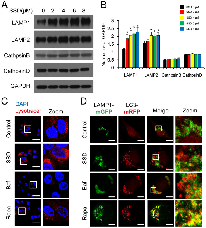
Figure 4.
SSD blocks autophagosome-lysosome fusion. (A) MDA-MB-231 cells were exposed to SSD for 24 h. The expression levels of LAMP1, LAMP2, cathepsin B and cathepsin D were analyzed using western blotting. (B) Semi-quantitative analysis of part A. Data are presented as the mean ± SD; n=3; *P<0.05 vs. SSD 0 µM. (C) MDA-MB-231 cells were exposed to 8 µM SSD, 20 nM Baf or 0.25 µM Rapa for 24 h and then stained with LysoTracker™ Red. Fluorescence was observed using a confocal laser microscope (scale bar=50 µm) (D) MDA-MB-231 cells were co-transfected with mRFP-LC3 and LAMP1-mGFP plasmids and then exposed to 8 µM SSD, 20 nM Baf or 0.25 µM Rapa for 24 h. The fluorescence of mRFP-LC3 and LAMP1-mGFP puncta was observed by confocal laser microscopy (scale bar=10 µm). SSD, saikosaponin D; Baf, bafilomycin A1; Rapa, rapamycin; LAMP, lysosome-associated membrane glycoprotein; GFP, green fluorescent protein; RFP, red fluorescent protein.

