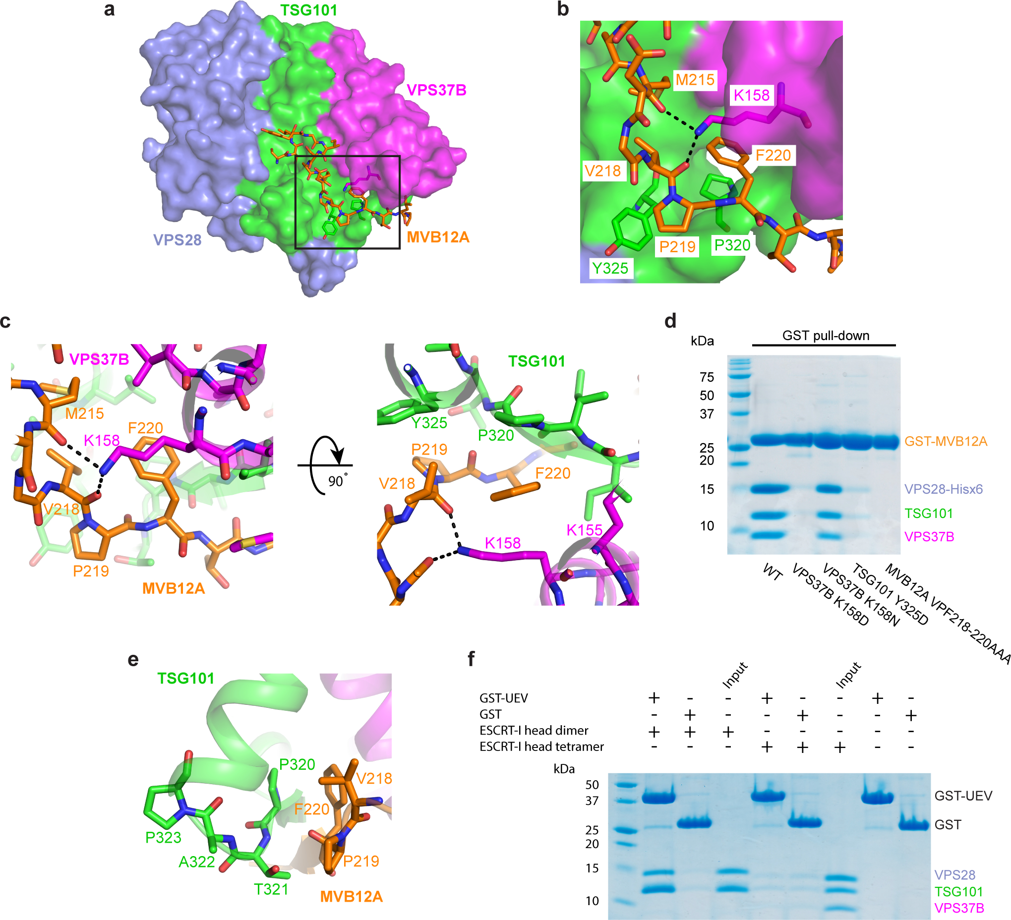
Fig. 2: MVB12A VPF motif is required for the interaction with the ESCRT-I head and occludes the TSG101 PTAP motif.

a, Surface representation of the human ESCRT-I head with MVB12A and other key residues shown as sticks. b, Enlargement of boxed region in (a) showing the conserved MVB12A VPF motif binding pocket. TSG101, VPS37B, MVB12A colored green, magenta and orange respectively. Selected hydrogen bonds are shown as dashed lines. c, Stick representation of the MVB12A VPF motif binding pocket. d, Mutant versions of the GST-tagged ESCRT-I head complex were expressed in E. coli. Lysate was incubated with glutathione beads and complex integrity analyzed by SDS-PAGE following multiple washes. e, Interface between TSG101 PTAP motif and MVB12A VPF motif. f, GSH beads were coated with GST-TSG101 UEV or GST bait and incubated with binary (TSG101–VPS28) or tetrameric (TSG101–VPS28 –VPS37B –MVB12A) ESCRT-I head. Beads were washed and complex formation analyzed by SDS-PAGE. The SDS-PAGE gels shown for the pull-down experiments in (d) and (f) represent two independent biological replicates. Uncropped images for panels (d) and (f) are in Supplementary Figure 1.
