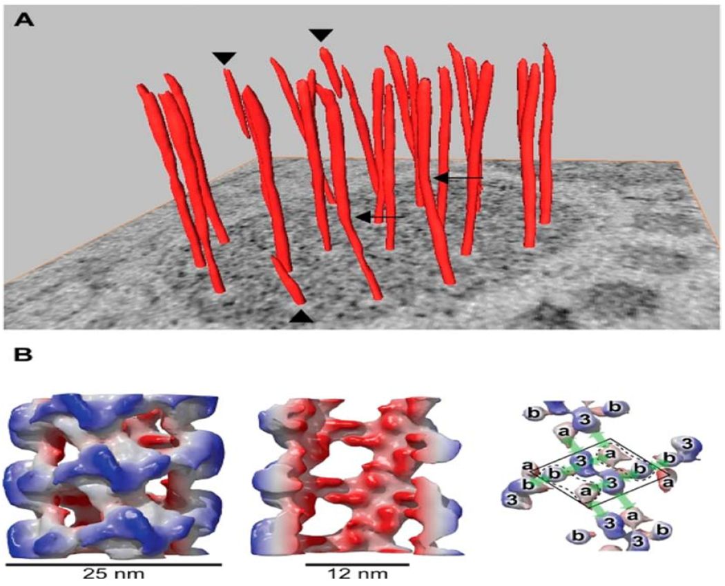
Figure 2:

Helical arrangement of VWF in tubular striations of WPB. (A) orderly twisting of the tubules within WPBs is well-illustrated on a tomographic slice. Several tubules (see arrowheads) stop halfway into the WPB and others (arrows) display kinks. In panel B, a reconstruction of VWF tubules is shown with an outside diameter of 25nm (left), cutaway view of the internal diameter of 12nm (middle) and different domains within the helix (right). From Valentijn KM et al. Blood 2011; 119: 5033-5043. With permission. VWF (Von Willebrand Factor), WPB (Weibel-Palade Bodies).
