
Figure 3. IL-22 signaling in the oral basal epithelial layer is required for protection against OPC.
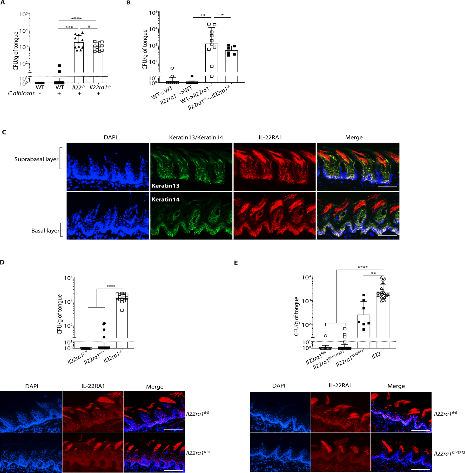
The indicated mice were subjected to OPC. A. Fungal burdens were assessed on day 5 p.i. Bars show geometric mean ± SD. Data were pooled from 2 independent experiments. B. BM from indicated donors was transferred into irradiated recipients. After 6–9 weeks, mice were subjected to OPC and fungal burdens assessed on day 5 p.i. Data were pooled from 2 experiments. C. Frozen sections from WT tongues were co-stained with DAPI and Abs against K13, K14 or IL-22RA1. Suprabasal and basal epithelial layers are indicated. Images are representative of a minimum of 3 sections. Size bar = 200 μm. D. Top: Fungal burdens were assessed on day 5 p.i. Data are pooled from 3 independent experiments. Bottom: IF staining of tongues from the indicated mice were co-stained with DAPI and α-IL-22RA1 Abs. Size bar = 200 μm. E. Top: All mice except Il22−/− were administered tamoxifen for 5 days prior to OPC, and fungal burden assessed on day 5 p.i. Bars show geometric mean ± SD. Bottom: Frozen sections from tongues from the indicated mice were co-stained with DAPI and α-IL-22RA1. Size bar = 200 μm. Data were pooled from 3 independent experiments and analyzed by ANOVA with Mann-Whitney U test.