
Figure 6. IL-22 licenses IL-17 signaling during OPC.
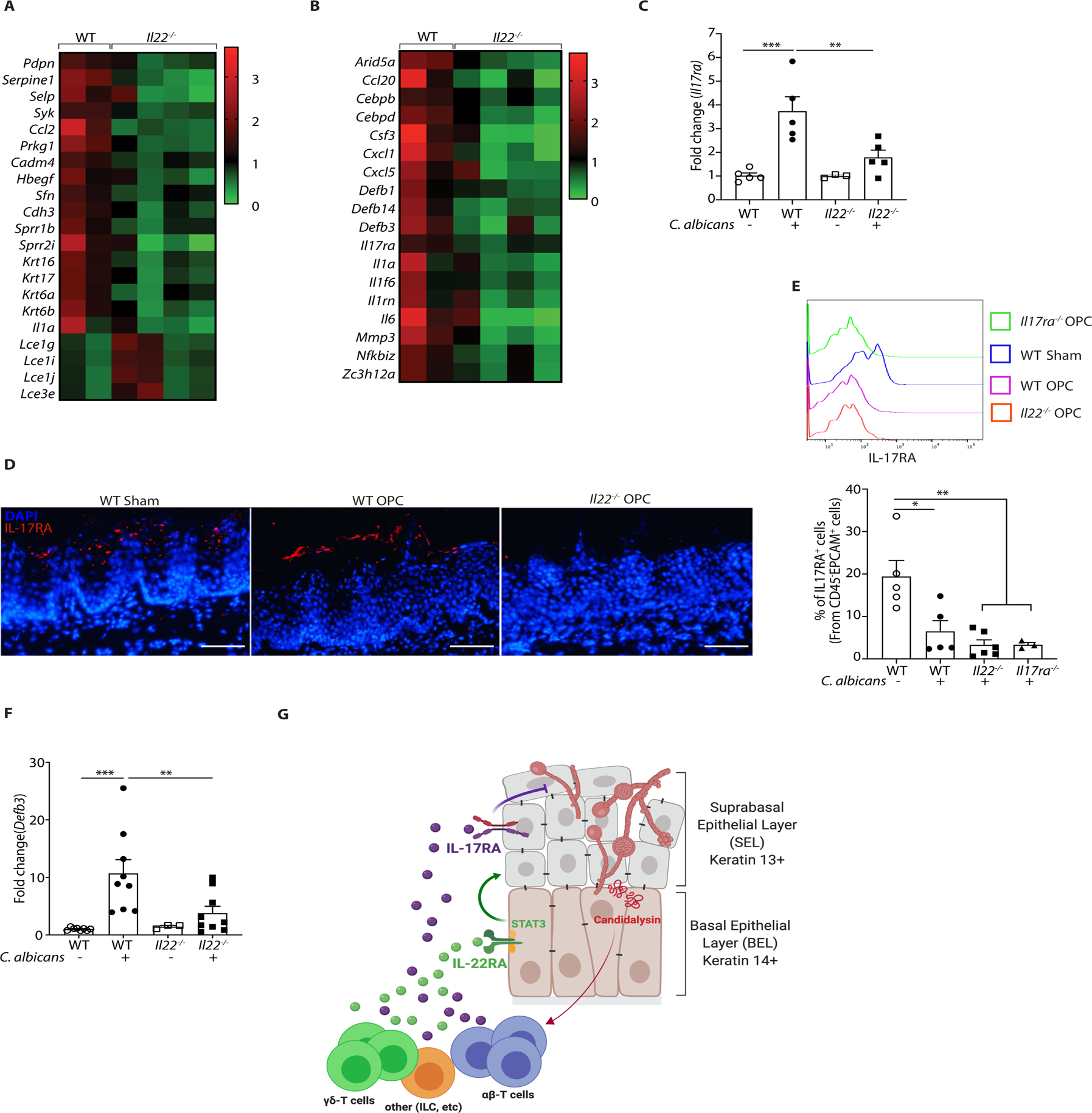
A. Heatmap of genes implicated in tissue repair, wound healing, keratinization and epithelial differentiation. B. Heatmap of IL-17 signature genes in OPC. C. qPCR of Il17ra expression normalized to Gapdh in tongue tissue from the indicated mice subjected to OPC and analyzed on day 2 p.i.. Data show mean ± SEM relative to sham-infected mice. Data analyzed by ANOVA or Student’s t-test. D. IF staining of IL-17RA and DAPI in the indicated mice on day 2 p.i. E. IL-17RA expression in CD45−EpCAM+ oral epithelial cells in WT or Il22−/− mice during OPC. Top: representative FACS histogram. Bottom: Pooled data from 2 independent experiments. Size bar = 200 μm. F. Expression of BD3 (Defb3) mRNA in tongue from WT or Il22−/− mice during OPC, normalized to Gapdh. Data analyzed by ANOVA or student’s t-test. G. Diagram of stratified oral epithelium during a first encounter with C. albicans. Fungal hyphae induces cellular damage and secrete the peptide candidalysin, which facilitates tissue invasion and activates innate IL-17- and IL-22-producing lymphocytes (see Refs (29, 61)). IL-17 was shown previously to act dominantly on K13+ cells of the suprabasal epithelial layer (SEL) (31). In contrast, IL-22/STAT3 promotes proliferation of the K14+ basal epithelial layer (BEL) that serves to restore the IL-17R-expressing SEL, thus maintaining IL-17-induced antifungal signals such as β-defensins and neutrophil responses that are required to mediate clearance of C. albicans. Diagram created on Biorender.com.