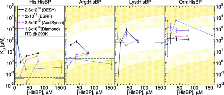
Figure 5.
Computed K D from V R curves in Fig. 4 ▸, plotted as a function of receptor concentration [HisBP] and total photon dose. Colours represent replicates at different total doses: (black) 2.6 × 10¹³ photons at DESY, (blue) 3 × 10¹² photons at ESRF, (red) 2.6 × 10¹³photons at Australian Synchrotron and (violet) 1.6 × 10¹⁴ photons at Diamond. Reference values from ITC are shown as dashed grey lines with error bars at the plot edges. Low confidence limits imposed by constant-receptor titrations are represented by light-yellow regions where K D lies outside one or two orders of magnitude of [HisBP]. For comparison, equivalent plots using other metrics are shown in Figs. S11–S13.

