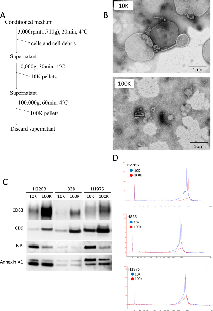
Fig 3. Association of long fragment DNA with extracellular vesicles (EVs) in lung cancer cell lines.
(A) Protocol for EV isolation using conditioned media of lung cancer cell lines. (B) Images of 10K and 100K pellets with a transmission electron microscope (TEM). (C) Western blotting analysis of 10K and 100K pellets. 5 μg of EV lysate was applied. We used antibodies to CD9 and CD63 as markers of small EVs (exosomes), and BiP and Annexin A1 as markers of large EVs. (D) DNA size distribution among 10K and 100K pellets.

