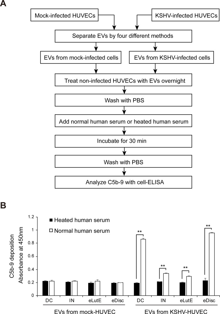
Fig 6. Activation of the complement system by EVs isolated from KSHV-infected cells using different methods.
(A) Schematic summary of the experimental process. Equal volumes of EVs separated by the four different methods from KSHV-infected HUVECs were applied to non-infected HUVECs with heated or normal human serum. The deposition of C5b-9 on the cells was analyzed by cell-ELISA. (B) The results of the cell-ELISA for C5b-9 in the non-infected HUVECs treated with separated EVs by various methods. DC: EVs separated using differential centrifugation. IN: EVs separated using the Invitrogen Total Exosome Isolation reagent. eLutE: EVs separated using the ExoLutE exosome isolation kit. eDisc: EVs separated using Exodisc of LabSpinner. Data are shown as the mean ± SD, n = 3, **p < 0.01.

