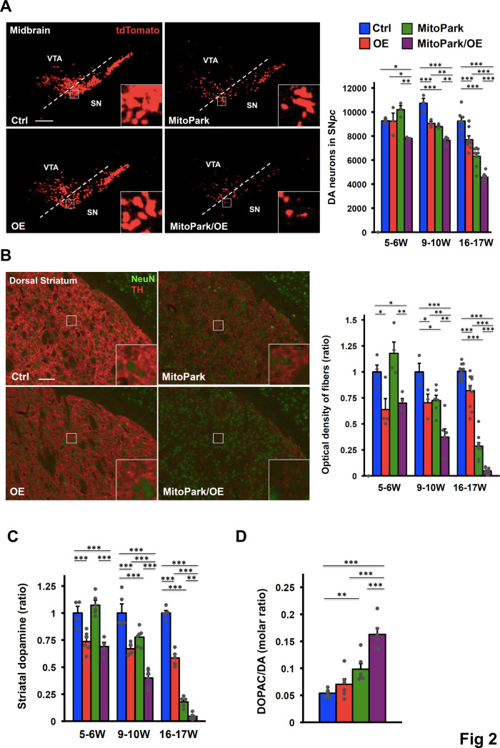
Fig 2. Elevated COUP-TFII expression contributes to progressive neurodegeneration of MitoPark mice.
(A) Representative images (left) of DA neurons in the ventral midbrain of 16- to 17-week-old Ctrl, OE, MitoPark, and MitoPark/OE mice detected by the tdTomato reporter and quantification (right) of DA neurons in the SNpc of mice at 5–6 (n = 3/group), 9–10 (n = 3-4/group), and 16–17 (n = 8/group) weeks of age (higher-power views in the insets). Scale bar, 200 μm. (B) Representative images (left) of DA axonal projections to the dorsal striatum of 16- to 17-week-old mice after double staining with anti-TH and anti-NeuN antibodies and quantification (right) of TH+ fibers of mice at 5–6 (n = 4/group), 9–10 (n = 3 or 6/group), and 16–17 (n = 10/group) weeks of age (higher-power views in the insets). Scale bar, 100 μm. (C) Relative total striatal dopamine levels of mice at 5–6, 9–10, and 16–17 weeks of age. n = 4-6/group. (D) Dopamine turnover, as measured by the ratio of the DA metabolite 3,4-dihydroxypheylacetic acid (DOPAC) to DA, in 9- to 10-week-old mice. n = 6/group. (A-D) *p < 0.05; **p < 0.01; ***p < 0.001. Mean ± SEM. One-way ANOVA Fisher’s LSD post hoc test. See also S2 Fig.

