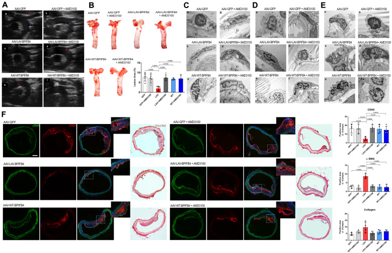
Figure 2.
LAV-BPIFB4 gene therapy hinders plaque formation, preserves vascular endothelium integrity, and reduces monocyte/macrophages infiltration. (A) Ultrasound scanning of aortic arch with epi-aortic vessels in AAV-treated ApoE−/− mice fed a high-fat diet. LAV-BPIFB4-treated mice did not have any plaques, whereas all those on the other treatments had calcified plaques (red arrows) and lipidic plaques (blue arrows). (B) Oil Red O staining and quantitative analysis of atherosclerotic lesion size in the aorta. Oil Red O staining was quantified using ImageJ software. One-way ANOVA followed by Tukey’s multiple comparisons test. Numbers above square brackets show adjusted P-values. (C–E) Representative micrographs of endothelial cells from aorta (C), mesenteric (D), and femoral (E) arteries: (a) Vascular endothelium of the AAV-GFP group showed cytosolic derangement (arrows) and broken plasma membranes (arrowheads); (b) these alterations were also evident in the group treated with the CXCR4 inhibitor. Diluted cytoplasm (arrow) and markedly condensed chromatin in the nucleus (N) was observed. (c) The endothelium was preserved by gene therapy with LAV-BPIFB4. (d) AMD3100 contrasted the benefit of LAV-BPIFB4, with the endothelial cells showing a diluted cytoplasm (arrows) and disruption of the plasma membrane (arrowheads); (e) The AAV-WT-BPIFB4-treated group presented with severely altered ultrastructure of the endothelium; (f) AMD3100 administration conferred additional ultrastructural damage, with endothelial cell detachment from the elastic membrane lamina (arrows). (F) Immunofluorescence staining of aortic arches from treated ApoE knockout mice, using the monocyte/macrophage marker CD68+, the smooth muscle cell marker αSMA, and Sirius Red staining to evaluate collagen (N = 4 per group). One-way ANOVA followed by Tukey’s multiple comparisons test. Numbers above square brackets show adjusted P-values.

