Figure 6.

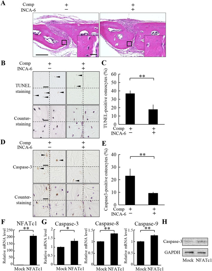
The induction of osteocyte apoptosis by compressive force loading is dependent on NFATc1. (A) Histological analysis of the parietal bones at 6 hours after application of compressive force was performed by H&E staining. INCA‐6 was injected s.c. into the parietal bone area prior to the application of compressive force. A magnified picture of the ROI is shown in the lower right corner of each picture. Scale bar = 100 μm, and 10 μm in insets. (B) TUNEL staining and subsequent counterstaining with hematoxylin of the parietal bones were performed. All four ROIs in a section in each group are shown. Arrowheads indicate TUNEL‐positive osteocytes. Comp = compressive force loading. Scale bar = 10 μm. (C) TUNEL‐positive ratio of osteocytes was calculated (four animals per group). **P < 0.01. (D) Caspase‐3 immunohistochemistry and subsequent counterstaining with hematoxylin of the parietal bones were performed. All four ROIs in a section in each group are shown. Arrowheads indicate caspase‐3‐positive osteocytes. Scale bar = 10 μm. (E) Caspase‐3‐positive ratio of osteocytes was calculated (four animals per group). **P < 0.01. (F–H) MLO‐Y4 cells were infected with a lentiviral vector encoding NFATc1 or an empty vector (mock). The expression of mRNA of NFATc1 (F), and caspase‐3, caspase‐8, and caspase‐9 (G) in MLO‐Y4 cells were analyzed by real‐time PCR. *P < 0.05, **P < 0.01; n = 3. (H) Cleaved caspase‐3 expression was analyzed by Western blot.
