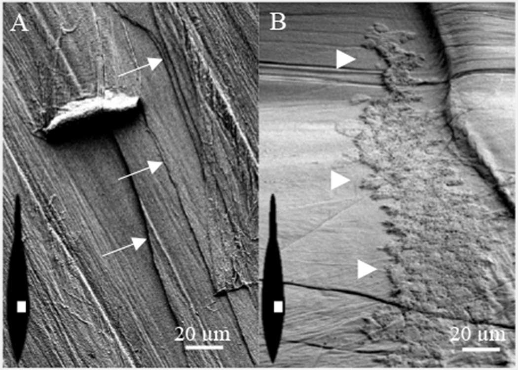
Figure 2.

S.E.M of pen surface. (A) The pen is composed of multiple layers identified by white arrows. (B) A region of the shell sac epithelium remains attached to the pen. White triangles identify the left perimeter. The illustrations within the images show the pen orientation (black) and region (white) used to collect the micrographs in A and B.
