
Figure 3.
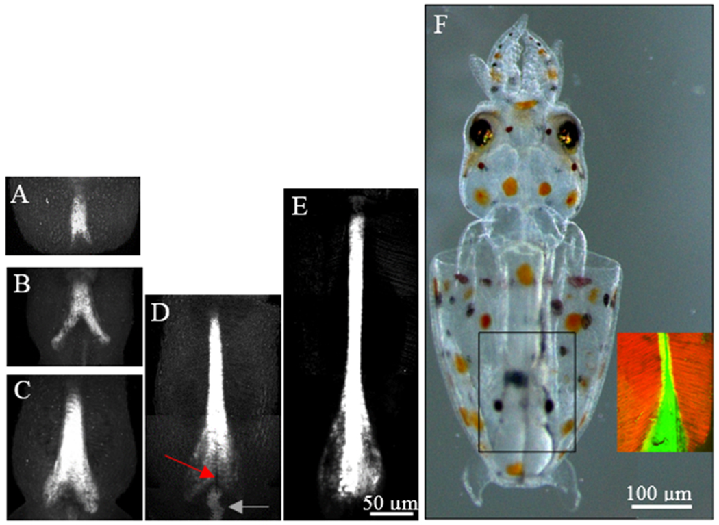
Elongation of the developing pen. Fluorescence imaging of Calcofluor white reveals pen development in fixed embryos. (A-C) Early stages show elaboration of chitin toward the anterior and the posterior vane. (D-E) In later stages, the anterior extends significantly longer while the vane expands and rounds off along the posterior edge. (F) In the hatchling, the actin filaments of the mantle (red) attach to the edges of the rachis and expanded vane of the pen (green) and do not completely encircle the pen.