Fig. 8.
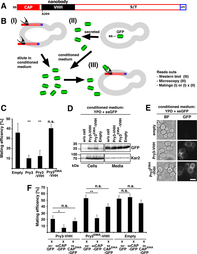
Cell wall association of the CAP domain results in a dose-dependent mating inhibition. (A) Schematic representation of cell-wall attached Pry3 internally tagged with the single-domain antibody VHH, which binds GFP. Pry3 is represented by a close red box for the CAP domain, a black box for the VHH internal tag, an open red box for the serine/threonine rich region, and a blue box for the GPI-anchor site. (B) Schematic representation of the nanobody-based assay system. (I) The nanobody-containing fusion protein is localized to the yeast cell wall (grey oval), exposed to the culture medium and thus can interact with GFP (green barrel) in the medium. (II) Cultivation of cell expressing a signal sequence (ss) containing GFP results in the secretion of ss-GFP into the culture medium. Tester strains (I) are then diluted in the conditional medium containing ss-GFP (III) and assayed for levels of the nanobody containing fusion protein by western blotting, its localization and functionality. (C) VHH-tagged Pry3 is functional in mating inhibition. Mating efficiency of cells containing an empty plasmid or overexpressing wild-type Pry3, Pry3-VHH, or Pry3E96A-VHH. Statistical analysis was performed as detailed in panel F. (D) Pry3-VHH and Pry3E96A-VHH are present at similar levels. Proteins from cells expressing Pry3-VHH, Pry3E96A-VHH or an empty vector as well as the conditioned culture medium containing ssGFP were analyzed by western blotting using an anti-GFP antibody. Equal signal intensity of GFP present in the conditioned medium used to cultivate cells expressing the different plasmids indicates that the availability of free GFP for binding to VHH is not limiting. Kar2 detection serves as loading control. (E) Pry3-VHH and Pry3E96A-VHH are localized to the yeast cell wall and bind GFP present in the conditioned medium. Cells containing an empty vector or expressing Pry3-VHH and Pry3E96A-VHH under the control of ADH1 promoter were cultivated in conditioned YPD medium containing free GFP for 4 h and analyzed by epifluorescence microscopy. BF, bright field; scale bar: 5 µm. (F) The CAP domain inhibits mating in a dose-dependent manner when localized to the cell wall. Quantitative mating assay of cells expressing the nanobody-containing wild-type Pry3, the E96A mutant version, or an empty plasmid. Cells were mated with strains expressing the secreted GFP (ss-GFP), secreted CAP domain (ss-CAP-GFP), or a E96A mutant version of the secreted CAP domain (ss-CAPE96A-GFP). (Welch t-test; *P-value <0.05; **P-value <0.01; n.s., not significant).

