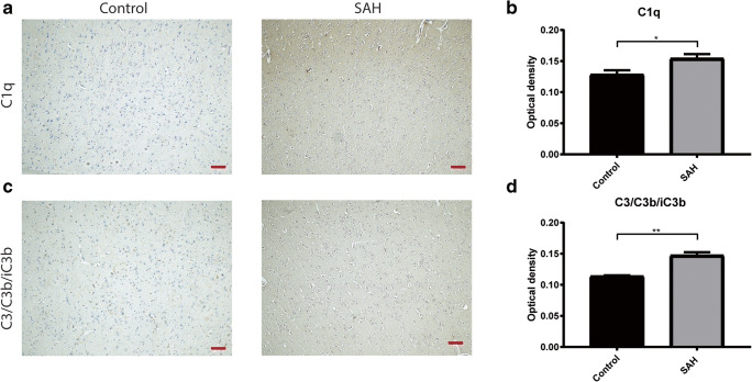
Fig. 1.
Complement expression in human autopsy brain sections. a Representative images of immunohistochemical staining of C1q on autopsy brain sections of a control patient, and a subarachnoid hemorrhage patient. b Average optical density measurements of C1q. c Representative images of immunohistochemical staining of C3/C3b/iC3b on autopsy brain sections of a control patient, and a subarachnoid hemorrhage patient. d Average optical density measurements of C3/C3b/iC3b. Scale bar: 100 μm, Student’s t test; *p ≤ 0.05; **p ≤ 0.01; mean ± SEM

