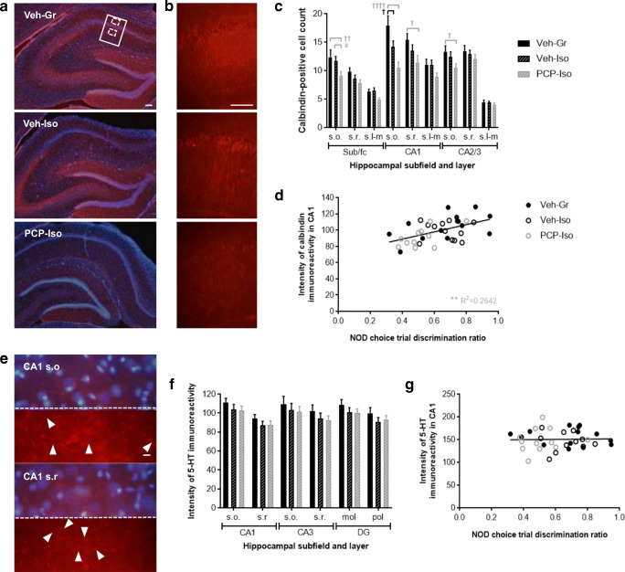
Fig. 5.
Impact of combined neonatal PCP and isolation rearing on hippocampal calbindin and 5-HT immunoreactivity in the dorsal hippocampus. Representative a, b calbindin immunoreactivity throughout a all subfields and b part of CA1 (indicated in a by a solid border), together with e higher magnification images of typical 5-HT immunoreactivity in CA1 strata oriens (s.o.) and radiatum (s.r.) locations (indicated in a by dashed borders). Red represents a, b calbindin or e 5-HT, and blue in a and the top portion of each image in e represents DAPI nuclear counterstain; the bottom portion of each image in e is a duplicate presented without the nuclear counterstain. Scale bars are equivalent to 100 μm a, b or 10 μm e. Mean ± SEM c number of calbindin-positive cells in s.o., s.r., and stratum lacunosum-moleculare (s.l-m) of the dorsal hippocampal subiculum/fasciola cinereum (sub/fc), CA1, and CA2/3, and f intensity of 5-HT immunofluorescence in s.o. and s.r. of dorsal hippocampal CA1 and CA3 and molecular (mol) and polymorphic (pol) layers of the dentate gyrus (DG), plus correlation analyses of d calbindin and g 5-HT immunofluorescence intensity in CA1 versus the NOD choice trial discrimination ratio (time exploring novel/total choice trial object exploration) following acute SB-399885. Male Lister hooded rats that received saline (1 ml/kg s.c.; Veh) or PCP (10 mg/kg) on PND 7, 9, and 11 were housed in groups (Gr; Veh only) or isolation (Iso; Veh and PCP) from weaning on PND 21, then underwent NOD on three occasions (PND 57–80) before tissue collection (PND 78–80), to receive vehicle (0.5% methylcellulose 1% Tween 80; 1 ml/kg i.p. 30 min before the familiarization trial), SB-399885 (10 mg/kg), or MMPIP (10 mg/kg) on separate test days in a pseudorandom order and serve as their own controls (n = 13–14 per neurodevelopmental condition). Calbindin immunoreactivity a was strongest in the dentate gyrus and stratum pyramidale, and remaining cells matched the distribution of GABA interneurons. The b intensity of calbindin immunoreactivity in CA1 and c number of calbindin-positive cells throughout dorsal hippocampal subfields and cell layers were both influenced by neurodevelopmental condition (P < 0.05 and P < 0.01, respectively), and although there were some reductions in single-hit Veh-Iso, these were much more extensive in PCP-Iso and correlated with d NOD performance following acute administration of SB-399885. Patterns of e 5-HT immunoreactivity were consistent with labeling of fine varicose axons (marked by arrowheads) but labeling intensity f was not influenced by neurodevelopmental condition and g did not correlate with NOD flowing SB-399885 administration. †P < 0.05 Veh-Iso and †P < 0.05; ††P < 0.01; ††††P < 0.0001 PCP-Iso versus Veh-Gr; #P < 0.05 PCP-Iso versus Veh-Iso (three-way repeated measures ANOVA with Tukey post hoc)

