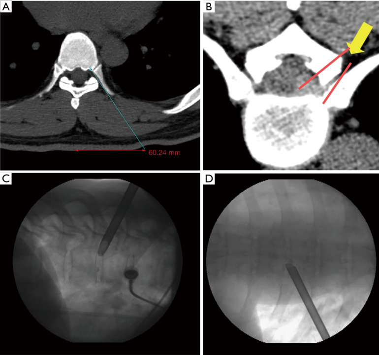
Figure 2.
Illustration of surgical trajectory. (A) Skin entry point planning on axial computed tomography scan by drawing an imaginary line from posterior annulus at the midpedicular level to lateral margin of facet joint; (B) imaginary line for foraminoplasty reaming of ventral and lateral aspect of superior facet; (C,D) placement of beveled working cannula on the posterior disc space through annular window at the posterior disc space—lateral view (C) and at the midpedicular level—AP view (D). AP, anteroposterior.

