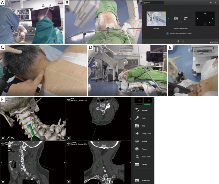
Figure 1.
Operating room set up. (A) This is an intraoperative view showing the surgeon looking at the attached monitors of both the endoscope and the neuronavigation system. (B) Reference setting, intraoperative computed tomography, and automatic registration. The red circles indicate the reflective calibration stickers that allow the navigation system to “see” the C-arm. (C) The patient is positioned adjusting the center line between the spinous process and the center of C arm rotation at which the green laser guidance shows midline. Operative setting for cervical pathologies. (D) This is the starting position of the C arm before acquiring the 3D scan. (E) The navigation pointer shows the entry to the pathology from the skin. (F) The navigation monitor in the hybrid operation room.

