
FIGURE 4.
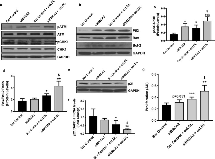
Oxidized LDL induces increased expression of DNA damage‐related molecules, p53, and related apoptotic molecules in BRCA2‐deficient endothelial cells. Proteins were extracted from HUVECs transfected either with scrambled control or siBRCA2 for 48 hr and then treated with oxLDL (100 μg/ml) for 24 hr to perform immunoblot for (a) p(phospho)ATM, ATM, pCHK1, and CHK1, (b) p53, Bax, and Bcl‐2, (e) p21. GAPDH was used as a loading control. Protein quantification for p53 (c) and for the Bax/Bcl‐2 ratio (d). (f) qPCR for p21. (g) Proliferation was evaluated in HUVECs transfected either with scrambled control or siBRCA2 for 48 hr and then treated with oxLDL for additional 24 hr. N = 3–4/group for immunoblot and qPCR in triplicates. Six‐wells/group for proliferation. *, ** and ***p < .05, 0.01 and 0.001 versus scrambled control, $ p < .05 versus scrambled control + oxLDL, p = .051 versus scrambled control