
Figure 3.
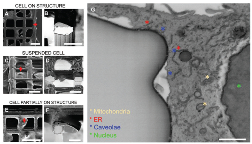
Cross sectioning of cells on ordered scaffolds. A) Top view of a cell growing on top of a square arm; B) 52° tilt view of cross section from position selected in (A); C) 52° tilt view of cell suspended and spreading between two arms; D) 52° tilt view of cross section from position selected in (C); E) top view of a cell partially attaching one arm and spreading over the perpendicular direction; F) 52° tilt view of cross section from position selected in (E); G) zoom-in of cross areas where cell located in (E),(F). (A), (C), (E) are acquired in secondary electrons mode. (B), (D), (F), (G) are acquired in backscattered mode and inverted. Scale bars: A) 50 μm, B) 20 μm, C) 30 μm, D) 20 μm, E) 50 μm, F) 20 μm, G) 1 μm.