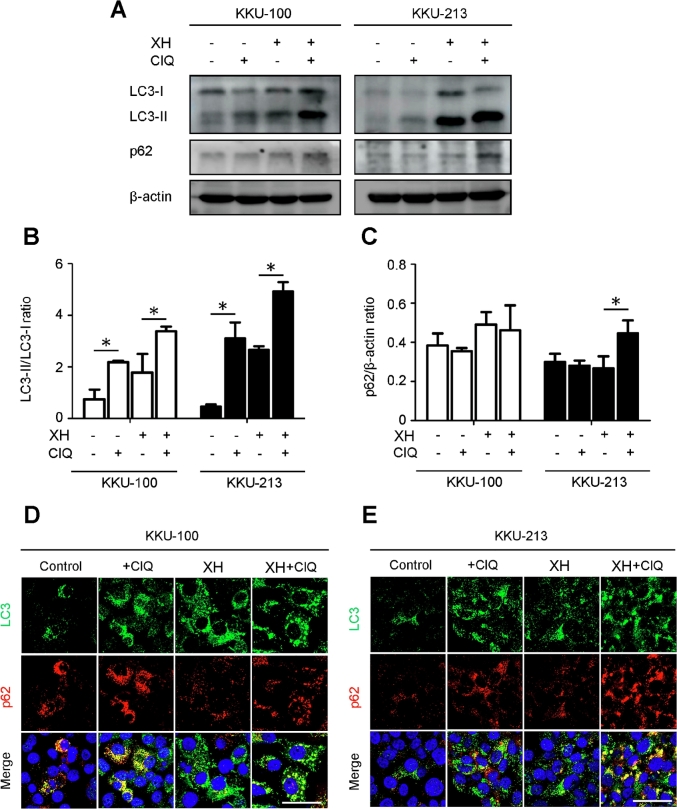
Fig. 2.
Xanthohumol induces autophagy in CCA cells. The expression of the autophagy markers LC3 and p62 was assessed by western blotting (A) and immuonofluorescence (D,E) in KKU-100 and KKU-213 CCA cell lines exposed to XH. Chloroquine (ClQ), which impairs autophagosome degradation, was added to discriminate between true induction of autophagy and block of the autophagy flux. Densitometry of relevant protein bands from three independent western blotting experiments is reported in panels B and C. The ratio of LC3-II/LC3-I indicates the neoformation of autophagosomes and the level of p62/β-actin indicates the autophagy degradation efficiency. Data are expressed as mean ± standard deviation of three independent experiments. Statistical significance between experimental conditions is indicated (* denotes a significant p-value of less than 0.05).

