Fig. 3.
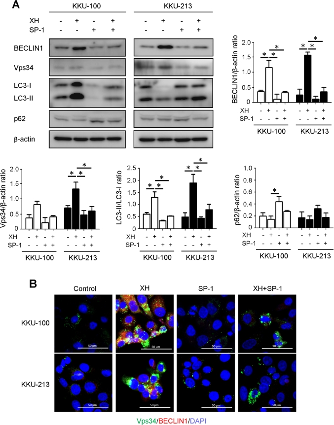
BECLIN 1 is an essential mediator of XH-induced autophagy. A: Western blotting analysis of BECLIN1, Vps34, LC3 and p62 expression in KKU-100 and KKU-213 exposed or not to XH in the absence or presence of Spautin-1 (SP-1) for 24 h. The filter was stripped and probed for β-Actin as a marker of protein loading. Densitometry analysis of the bands from three independent western blotting is reported in the histogram. *p < 0.05 compared to control. (B) Double immunostaining for BECLIN1 (red fluorescence) and Vps34 (green fluorescence) in CCA cells treated with XH ± SP-1 as indicated. Nuclei were counterstained with Hoechst 33342 (blue fluorescence). Scale bar = 20 μM; magnification = 63x. Representative images of three independent experiments. Data in this figure demonstrate that SP-1-induced degradation of BECLIN-1 abrogates autophagy induced by XH.

