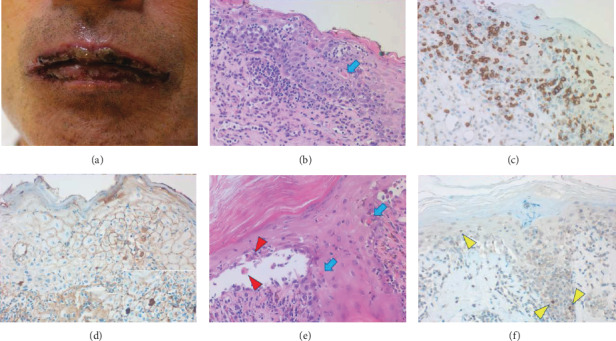
Figure 1.

Skin biopsy of the lip and abdomen. The lips grossly reveal extensive erosion (a). Microscopically, both the spongiotic epidermis with graft-versus-host disease-like appearance and upper dermis are infiltrated by small lymphocytes (b, HE). Blue arrow indicates a Civatte body. The lymphocytes are predominantly immunoreactive for CD8 (c). After proteinase-1 digestion of the paraffin section, IgG deposition along the plasma membrane of the involved keratinocytes is proven (d). IgM is also deposited (inset). Dermal IgG positivity represents the normal distribution of IgG in tissue fluid. The abdominal skin exhibits pemphigus vulgaris-like interface blister formation. Acantholytic keratinocytes are indicated by red arrowheads, and Civatte bodies are shown by the blue arrows (e, HE). Yellow arrowheads indicate cleaved caspase-3 immunoreactivity in apoptotic keratinocytes (f).
