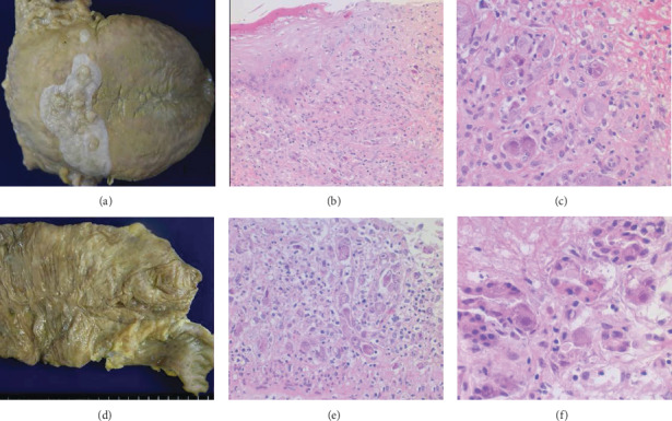
Figure 2.

Opportunistic CMV infection in the digestive tract. The tongue is extensively eroded: the white-colored mucosa around the vallate papillae remains intact (a). Glossal erosion adjacent to intact squamous mucosa reveals infection of CMV (b, HE), and the high-powered view clarifies CMV infection in the endothelial cells (c, HE). Multifocal erosions are formed in the mucosae of the terminal ileum and cecum (d). The endothelial cells of the eroded cecal mucosa are heavily infected by CMV, and crypt epithelial cells are lost (e). CMV infection in the pancreas has provoked fat necrosis (f).
