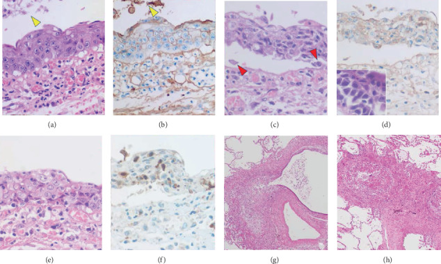
Figure 4.

PNP-related bronchial/bronchiolar lesions. The regenerative bronchial mucosa focally with basal intercellular vesicle formation is associated with IgG deposition on the plasma membrane (a: HE, b: IgG immunostaining after proteinase-1 digestion). Intraluminal cellular debris is also labeled for IgG (yellow arrowheads). The stromal labeling represents endogenous IgG distributed in the tissue fluid. (c, HE and d, IgG) Interface blister formation with IgG deposition on the epithelial cells is demonstrated. Acantholytic cells are indicated by red arrowheads. Inset demonstrates acantholytic change of the bronchial mucosa (HE). Another part of the disorganized bronchial mucosa shows clustering of apoptotic cells immunoreactive for cleaved caspase-3 (e: HE, f: cleaved caspase-3 immunostaining). Microscopic features of BO are observed in the peripheral lung. Mucosal erosion-associated exudation has provoked secondary luminal dilatation (g, HE) and luminal obstruction (h, HE). Peribronchiolar fibrosis is noted.
