Figure 3.
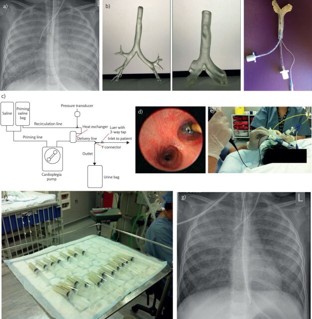
Whole-lung lavage in a 2-year old girl with PAP due to a homozygous CSR2RB mutation. a) Chest radiograph prior to lavage. There is diffuse, bilateral nonspecific shadowing. The appearances are not specific for PAP. b) Three-dimensional model of trachea with two endotracheal tubes in situ. The model, printed from HRCT images, allows pre-procedure planning of ventilation, bronchial blocker and lavage strategy in patients in whom a double-lumen tube cannot be passed. c) Schematic diagram of the semiautomated circuit used for whole-lung lavage in smaller patients. d) Bronchoscopic view of the bronchial blocker in the left main bronchus, prior to instilling lavage fluid into the left lung. e) Whole-lung lavage. The bronchoscopist is carefully checking the position of the bronchial blocker, while a second operator is, in this case, using a syringe to perform the lavage. Note the creamy-coloured fluid in the aspirating syringe, typical of PAP. f) Serial aliquots of lavage fluid. Note that as the lavage has proceeded, the fluid becomes clearer. g) Chest radiograph following right sided lung lavage in the same child as in part a. There is substantial clearing of the changes in the right middle and lower lobes; the right upper lobe could not be lavaged because it was impossible to obtain a stable occlusion position in the right main bronchus and the bronchial blocker was placed in the right bronchus intermedius.

