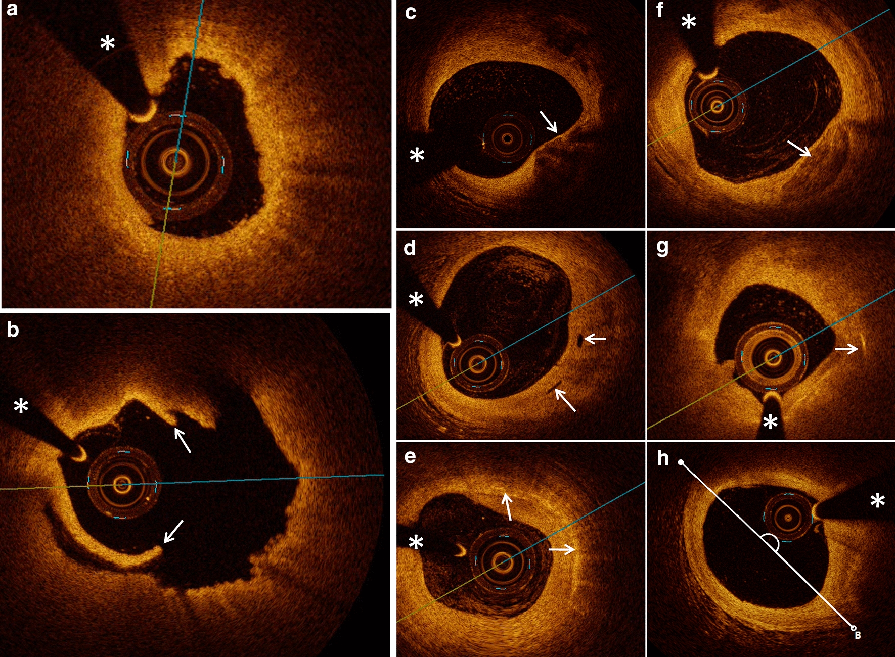
Fig. 1.

Typical optical coherence tomography (OCT) images of plaque characteristics. a A typical image of a patient with STEMI and plaque erosion characterized as intact and rouHbA1c intima attached with small white thrombus. b A typical image of a patient with STEMI and plaque rupture characterized as ruptured intima (arrow) backed with a big cavity. c–h Representative OCT images, including TCFA, micro-channel, macrophage accumulation, spotty calcification, cholesterol crystal, and lipid core measured with the lipid arc, are presented in turn (arrows). The asterisk marks the position of the OCT guide wire
