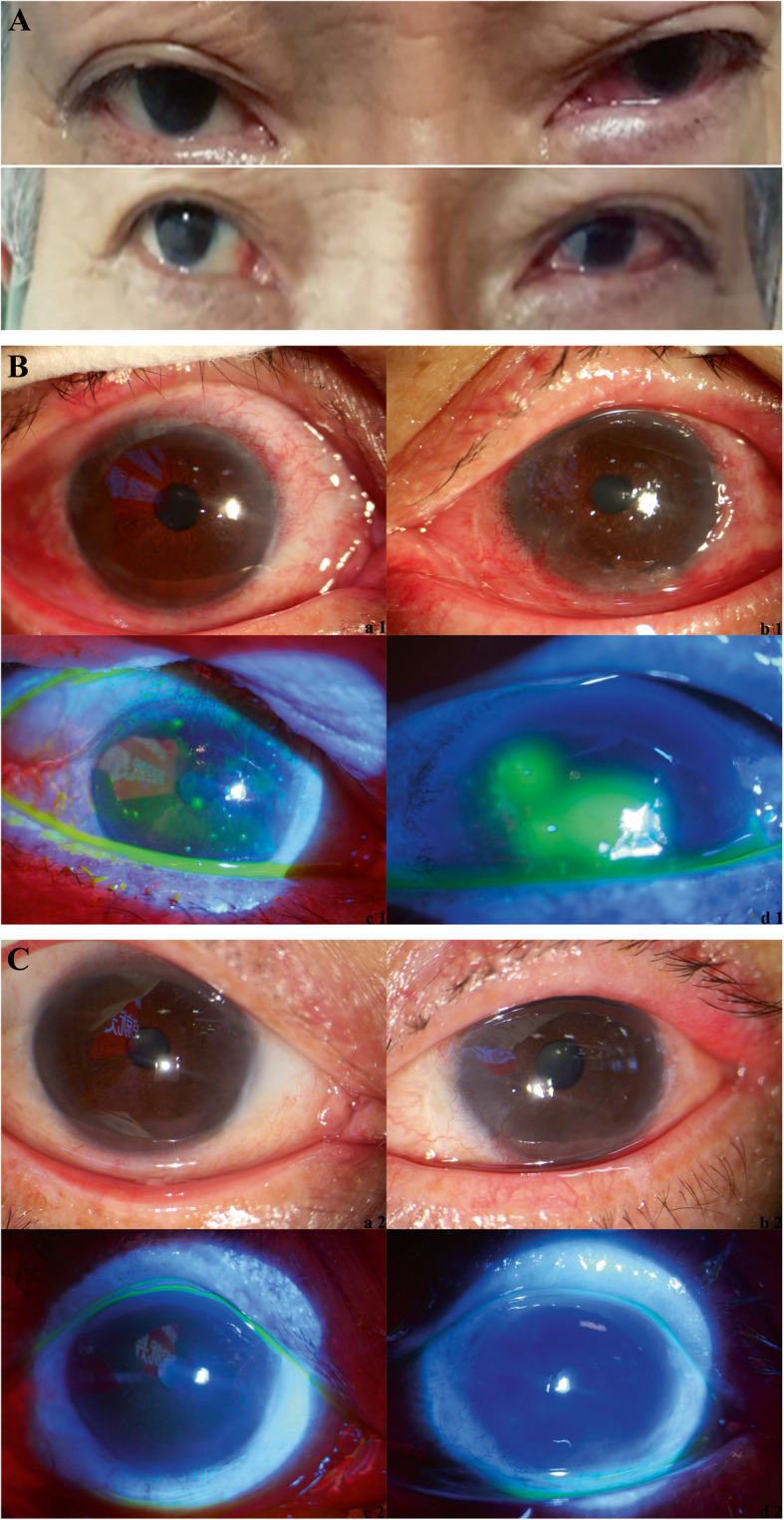
Fig. 2.

Eye images of the patients. a Image taken on 16 February 2020. b Slit lamp images taken on 1 March 2020. Panels (a1) and (c1) show the right eye and (c1) shows the right eye stained with fluorescein sodium and visualized under a cobalt blue light. Panels (b1) and (d1) show the left eye and (d1) shows the left eye stained with fluorescein sodium and visualized under a cobalt blue light. c Slit lamp images taken on 8 March 2020. Panels (a2) and (c2) show the right eye, and (c2) shows the right eye stained with fluorescein sodium and visualized under a cobalt blue light. Panels (b2) and (d2) show the left eye, and (d2) shows the left eye stained with fluorescein sodium and visualized under a cobalt blue light.
