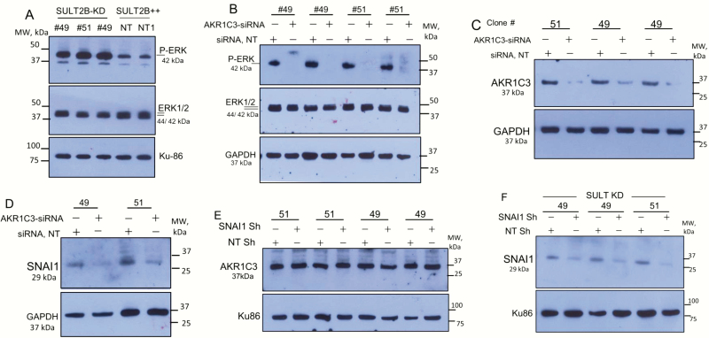
Figure 5.
ERK1/2 activation in SULT2B-KD cells, and the role of AKR1C3 in ERK activation and SNAI1 induction. (A) Phospho-ERK1/2 in KD clones (#49, #51) and non-KD clone. Clone #49 and NT/NT1 non-targeting clone were assayed in biological replicates. (B) Loss of ERK1/2 activation in AKR1C3-silenced SULT2B KD cells. Clones #49 and #51 were assayed in biological replicates. (C) AKR1C3 silencing by siRNAs in SULT2B KD clones. (D) Reduced SNAI1 expression in AKR1C3-silenced KD clones. E) AKR1C3 levels in SNAI1 knockdown and SNAI1 intact SULT2B KD cells. (F) SNAI1 levels in SULT2B KD cells treated with SNAI1 shRNA lentivirus or scramble shRNA lentivirus.

